Home » Without Label » Back Of Neck Anatomy - Why Neck Training Should Be A Priority For Athletes / Anatomical principles underlying cranial nerve lesions;
Back Of Neck Anatomy - Why Neck Training Should Be A Priority For Athletes / Anatomical principles underlying cranial nerve lesions;
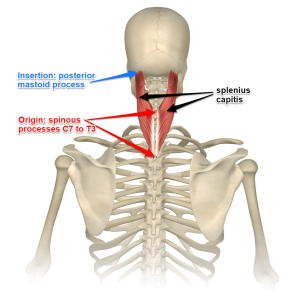
Back Of Neck Anatomy - Why Neck Training Should Be A Priority For Athletes / Anatomical principles underlying cranial nerve lesions;. From the sides and the back of the neck, the splenius capitis inserts onto the head region, and the splenius cervicis extends onto the cervical region. The neck begins at the base of the skull and connects to the thoracic spine (the upper back). The anterior muscles of the neck facilitate swallowing and speech. A collection of anatomy notes covering the key anatomy concepts that medical students need to learn. & post triangles •this is a large muscle in the anterior neck •starting at the manubrium of the sternum and the clavicle and going to the mastoid process of the.
Clinically, surface anatomy is used to split the neck into anterior and posterior triangles which provide clues as to the location of specific structures. The levator scapulae muscle is attached at the top four cervical vertebrae (c1 to c4) and runs down the side of the neck to attach at the top of the shoulder blade (scapula). This article describes the anatomy of the head and neck of the human body, including the brain, bones, muscles, blood vessels, nerves, glands, nose, mouth, teeth, tongue, and throat. Start studying anatomy of neck & back. Click now to study the muscles, glands and organs of the neck at kenhub!
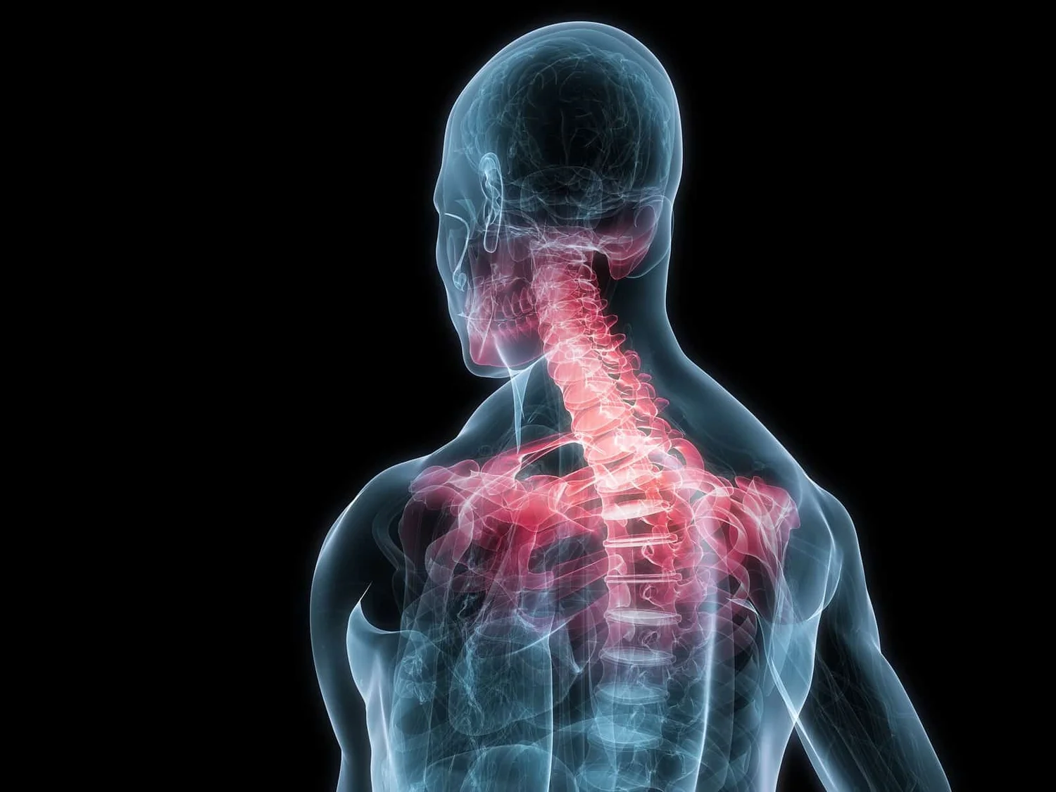
The Link Between Posture And Chronic Neck And Upper Back Pain Back Pain And Headache Specialist Burke Va Nova Headache Chiropractic Center from images.squarespace-cdn.com This mri neck axial cross sectional anatomy tool is absolutely free to use. The levator scapulae muscle is attached at the top four cervical vertebrae (c1 to c4) and runs down the side of the neck to attach at the top of the shoulder blade (scapula). The anterior muscles of the neck facilitate swallowing and speech. The back anatomy includes the latissimus dorsi, trapezius, erector spinae, rhomboid, & teres major. The cervical spine supports the weight and movement of your head and pro. This entry was posted in anatomy by admin. Start studying anatomy of neck & back. Our neck is where we find the seven cervical vertebrae, with c7 (the seventh cervical vertebra) meeting t1 (the first thoracic vertebra) at the base of the neck.
This entry was posted in anatomy by admin.
Learn vocabulary, terms and more with flashcards, games and other study tools. The cervical spine supports the weight and movement of your head and pro. From the sides and the back of the neck, the splenius capitis inserts onto the head region, and the splenius cervicis extends onto the cervical region. We've largely focused on the physical aspect of our spinal anatomy in this series. Discography is a diagnostic procedure the back experts at the southeastern spine institute (ssi) use to determine if any of your intervertebral discs are the primary cause of your back pain. Beneath the integument the back of neck presents in the median plane the ligamentum nuchae, which is a triangular fibrous sheet and represents upward continuation of supraspinous ligament. Of the head, neck and vertebral column; Neck, in land vertebrates, the portion of the body joining the head to the shoulders and chest. It runs from the neck to the upper back. The suprahyoid muscles originate from above the hyoid bone in the chin region. « back show on map ». See more ideas about anatomy, anatomy and physiology, muscle anatomy. Clinically, surface anatomy is used to split the neck into anterior and posterior triangles which provide clues as to the location of specific structures.
This article describes the anatomy of the head and neck of the human body, including the brain, bones, muscles, blood vessels, nerves, glands, nose, mouth, teeth, tongue, and throat. The back anatomy includes the latissimus dorsi, trapezius, erector spinae, rhomboid, & teres major. When most people mention their back, what they are actually referring to is their spine. The splenius muscles originate at the midline and run laterally and superiorly to their insertions. The cervical spine is the top part of the spine.
Structure And Function Of The Cervical Spine Physiopedia from www.physio-pedia.com Discography is a diagnostic procedure the back experts at the southeastern spine institute (ssi) use to determine if any of your intervertebral discs are the primary cause of your back pain. This is often a result of incorrect posture. This mri neck axial cross sectional anatomy tool is absolutely free to use. Head and neck anatomy is important when considering pathology affecting the same area. Last time we learned that the trapezius makes the back wall of the neck. The structure is, of course, an important part of the conversation. Learn about these muscles, their locations & functional the traps are quite a complex set of muscles. The sections below will cover these elements in more detail.
Head and neck anatomy is important when considering pathology affecting the same area.
It provides images in the axial and coronal planes so that the user can study and learn anatomy. Head and neck anatomy is important when considering pathology affecting the same area. It is made up of bones, discs the neck is connected to the upper back through a series of seven vertebral segments. Sternocleidomastoid muscle (main muscle in the front of the neck) thyroid gland The back anatomy includes the latissimus dorsi, trapezius, erector spinae, rhomboid, & teres major. It runs from the neck to the upper back. In radiology, the 'head and neck' refers to all the anatomical structures in this region excluding the central nervous system, that is, the brain and spinal co. It is made up of bones discs muscles ligaments nerves and tendons. Learn everything about the neck anatomy with this topic page. Learn vocabulary, terms and more with flashcards, games and other study tools. But, what the neck is all. Some important structures contained in or passing through the neck include the seven cervical vertebrae and enclosed spinal cord, the jugular veins and carotid arteries, part of the esophagus, the larynx. This entry was posted in anatomy by admin.
They control the scapulae (shoulder blades), which play a role in shrugging, neck movement, head. The back comprises the spine and spinal nerves, as well as several different muscle groups. 3d video tutorials and interactive modules on the anatomy of the back including anatomy of the musculature, vertebral column, joints and ligaments. The sections below will cover these elements in more detail. Start studying anatomy of neck & back.
Understanding The Spinotransversales The Superficial Intrinsic Muscles Of The Back from www.nfpt.com They control the scapulae (shoulder blades), which play a role in shrugging, neck movement, head. 3d video tutorials and interactive modules on the anatomy of the back including anatomy of the musculature, vertebral column, joints and ligaments. Use the mouse scroll wheel to move the images up and down alternatively use the tiny arrows (>>) on both side of the image to move the images. This article describes the anatomy of the head and neck of the human body, including the brain, bones, muscles, blood vessels, nerves, glands, nose, mouth, teeth, tongue, and throat. We've largely focused on the physical aspect of our spinal anatomy in this series. A collection of anatomy notes covering the key anatomy concepts that medical students need to learn. Many in the neck help to stabilize or move the head. The sections below will cover these elements in more detail.
D) demonstrate sound knowledge of the surface/living and radiological anatomy.
Our neck is where we find the seven cervical vertebrae, with c7 (the seventh cervical vertebra) meeting t1 (the first thoracic vertebra) at the base of the neck. Start studying anatomy of neck & back. How to view the anatomical labels. Discography is a diagnostic procedure the back experts at the southeastern spine institute (ssi) use to determine if any of your intervertebral discs are the primary cause of your back pain. The levator scapulae muscle is attached at the top four cervical vertebrae (c1 to c4) and runs down the side of the neck to attach at the top of the shoulder blade (scapula). 3d video tutorials and interactive modules on the anatomy of the back including anatomy of the musculature, vertebral column, joints and ligaments. Neck, in land vertebrates, the portion of the body joining the head to the shoulders and chest. The splenius muscles originate at the midline and run laterally and superiorly to their insertions. Your neck is like no other part of the vertebral spinal column and enables your head and neck a wide range of motion. Learn vocabulary, terms and more with flashcards, games and other study tools. The cervical spine supports the weight and movement of your head and pro. Want to learn more about it? Integrates anatomy and physiology of cells, tissues, organs, the systems of the human body, and mechanisms responsible for homeostasis.